patandfamBlogModules

Differentials

Written and reviewed by Nigerian physicians and medical educators

Tuberculosis isn't the only disease that forms granulomas or enlarges a lymph node. Know what separates it from these.

Sarcoidosis
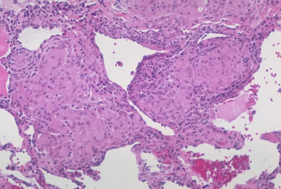
Lung tissue with several compact pink granulomas made of pale epithelioid cells and large multinucleate giant cells, ringed by small dark lymphocytes. No necrotic centre.
Sarcoidosis. Non-necrotising granulomas with many multinucleate giant cells, and no caseation.Yale Rosen, Atlas of Pulmonary Pathology · CC BY-SA 2.0 · reproduced unmodified
What looks the same
Granulomas without caseation. It also raises calcium through vitamin D, just as tuberculous granulomas do.
What separates it
Not the slide alone, because tuberculous granulomas can lack caseation too. Stain for acid-fast bacilli whenever you see granulomas.

Lymphoma
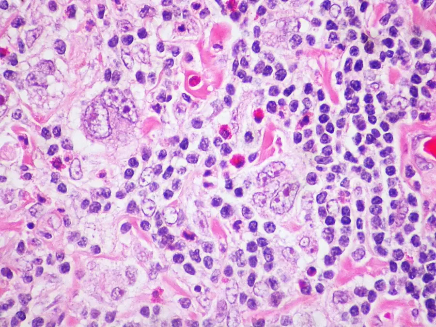
High power view of many small dark lymphocytes, scattered bright red eosinophils, pink collagen bands, and a few very large cells with big pale nuclei and prominent nucleoli.
Hodgkin lymphoma, nodular sclerosis type. Large atypical Reed-Sternberg-type cells among small lymphocytes, eosinophils and bands of collagen. No granulomas and no caseation.Yale Rosen, Atlas of Pulmonary Pathology · CC BY-SA 2.0 · reproduced unmodified
What looks the same
An enlarged lymph node. On a peripheral node biopsy in Nigeria, tuberculosis and the lymphomas are the two leading diagnoses.
What separates it
Caseation points to tuberculosis. Reed-Sternberg cells point to lymphoma. The gross specimen cannot decide it.

Foreign body reaction
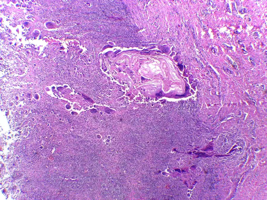
Low power view of pale pink flakes of keratin surrounded by large multinucleate giant cells and a dense sheet of small inflammatory cells.
Foreign body granuloma: a granulomatous reaction to keratin, with foreign body giant cells and chronic inflammatory cells.Department of Pathology, Calicut Medical College · CC BY-SA 4.0 · reproduced unmodified
What looks the same
Multinucleate giant cells.
What separates it
In a foreign body giant cell the nuclei are scattered at random through the cytoplasm. In a Langhans giant cell they sit in a horseshoe at the edge.
Study this in the moduleThe same topic, with pictures to explore, quick checks and your notes.Practice questionsFree accountExam-style questions, practical stations and a spoken viva on Tuberculosis.FlashcardsCards from every specialty, and any key facts you've saved.

Every topic in Tuberculosis