Pathogenesis
Key facts
- Inhaled bacilli survive in alveolar macrophages because sulfatides block phagosome-lysosome fusion.
- The Ghon focus and its caseating node form the Ghon complex; once calcified, it's called the Ranke complex.
- Primary disease sits low in the upper lobe or high in the lower lobe; secondary disease goes to the apex.
How an inhaled organism survives, sets off the immune response, and leaves a lesion behind.
Pathogenesis of tuberculosis, step by step
- Inhalation and phagocytosis. Droplet nuclei are small enough to slip past the mucus of the upper airway and reach an alveolus, each carrying only a few bacilli. They land at the bottom of the upper lobe or the top of the lower lobe, usually close to the pleura. An alveolar macrophage phagocytoses them.
- The organism survives phagocytosis. Sulfatides in the wall inhibit the phagosome from fusing with the lysosome that carries the bactericidal enzymes. Phagocytosed bacteria are not destroyed. They multiply within the macrophage, which then carries them through lymphatics and blood to distant sites.
- Sensitisation in the draining node. Bacilli, free or within phagocytes, travel via lymphatics to the regional nodes, which also often caseate. Macrophages present antigen to T helper cells, and sensitised T cells multiply and enter the circulation in search of the organism.
- Activated macrophages destroy tissue as well as bacteria. Sensitised T cells returning to the lesion activate the macrophages, which acquire epithelioid morphology and attack the bacteria. The attack destroys lung tissue at the same time. The necrotic centre is granular and cheese-like: caseous necrosis. Some epithelioid cells fuse into Langhans giant cells.
- Ghon focus, Ghon complex, Ranke complex. The parenchymal lesion is the Ghon focus, a grey-white area of consolidation one to one and a half centimetres across. Combined with the caseating node that drains it, it is the Ghon complex. Cell-mediated immunity controls the infection in most people, the complex fibroses and calcifies, and the healed pair is the Ranke complex.
- Or dissemination. Where containment fails, the apical caseous focus expands and erodes into a bronchus, evacuating its centre and leaving a cavity. Affected patients now produce sputum containing bacilli, and disease disseminates along the airways.
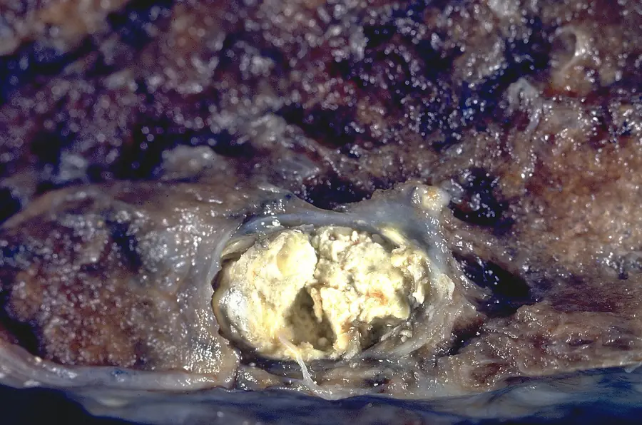
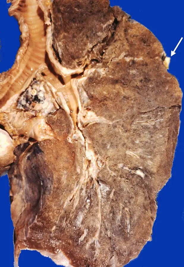
At autopsy you find a calcified subpleural nodule and a calcified hilar node in someone who was never diagnosed with tuberculosis. What happened to this person?
They were infected, but their cell-mediated immunity contained it. That's what happens in most people. The parenchymal focus and the node that drained it both fibrosed and calcified, leaving the Ranke complex. Organisms may still have been alive inside it. Had their resistance failed at any point, the lesion would have reactivated.