patandfamBlogModules

Morphology: gross

Written and reviewed by Nigerian physicians and medical educators

What to look for on the lung specimen.
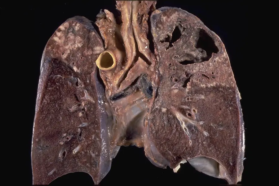
Cut lung showing a large irregular cavity in the upper zone.
Cavitary tuberculosis. Necrotic material has emptied into a bronchus, leaving a cavity open to the airway.Yale Rosen, Atlas of Pulmonary Pathology · CC BY-SA 2.0 · reproduced unmodified
  1. The cavity Erosion of an apical caseous focus into a bronchus empties the necrotic centre, leaving a ragged, irregular cavity lined by caseous material. The sputum now carries bacilli, so a patient with a cavity is highly infectious.
  2. The cavity wall Ragged, firm and poorly walled off by fibrous tissue. Erosion of blood vessels in this wall produces haemoptysis.
  3. Multiple areas of softening The upper zones are riddled with grey-white caseation and several separate areas of softening and cavitation, rather than one clean defect. This is coalescence of nodules over months.
  4. The lower zones Congested but not cavitated. Secondary disease localises to the apex of one or both upper lobes. This may relate to the high oxygen tension there, since the organism is an obligate aerobe.
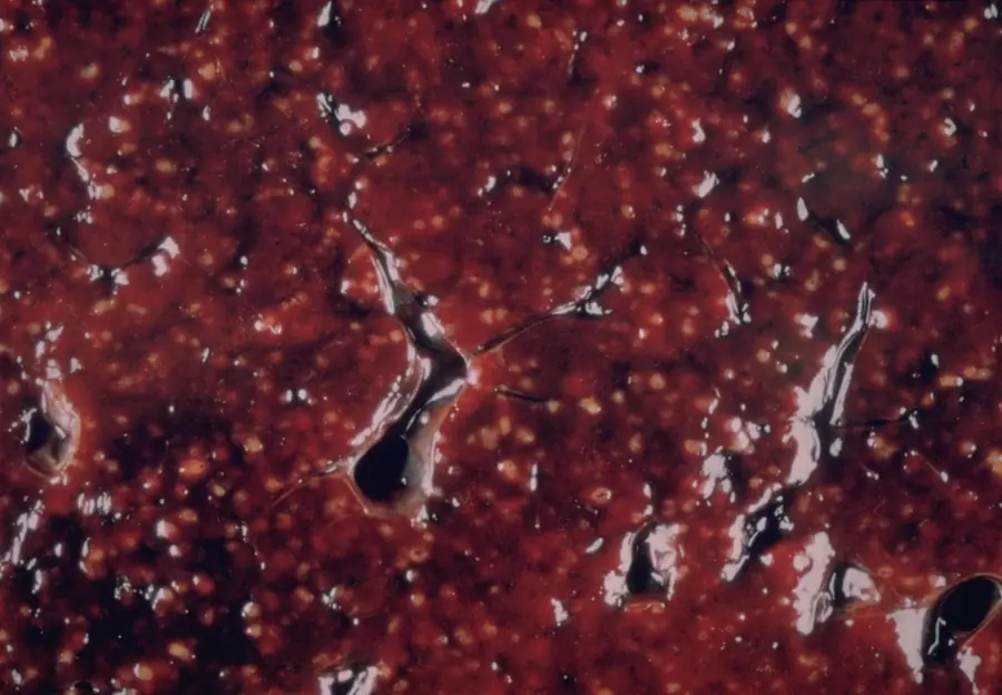
Cut spleen studded with countless small pale nodules of even size.
Miliary tuberculosis involving the spleen. Innumerable nodules, all of similar size, which is the discriminating feature.Yale Rosen, Atlas of Pulmonary Pathology · CC BY-SA 2.0 · reproduced unmodified
Study this in the moduleThe same topic, with pictures to explore, quick checks and your notes.Practice questionsFree accountExam-style questions, practical stations and a spoken viva on Tuberculosis.FlashcardsCards from every specialty, and any key facts you've saved.

Every topic in Tuberculosis