Morphology: histology
Key facts
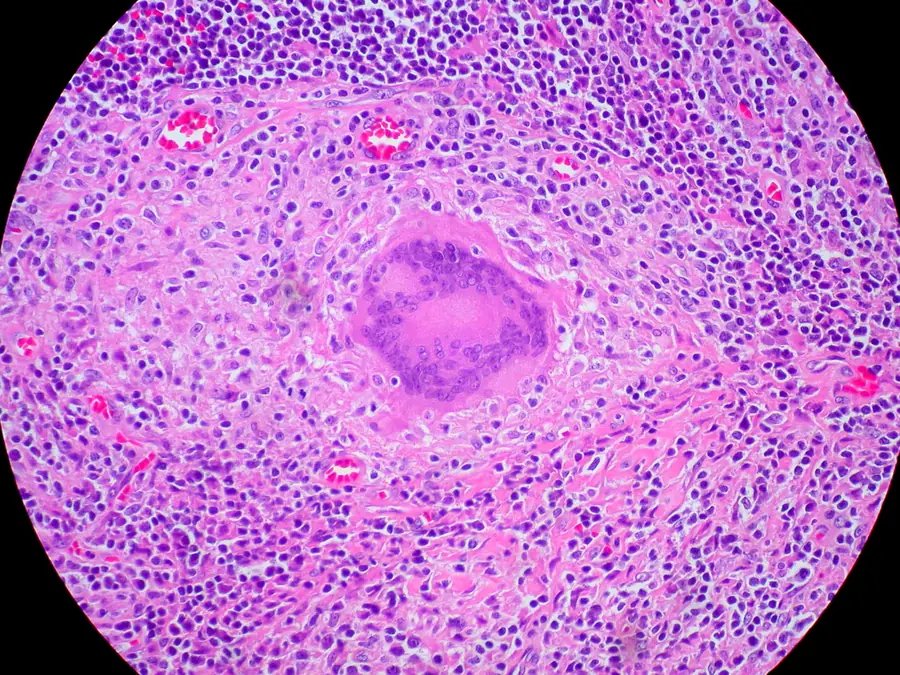
- Caseation sits at the centre, ringed by epithelioid and giant cells, then lymphocytes and fibroblasts.
- A Langhans giant cell has its nuclei in a horseshoe at the edge; a foreign body giant cell's nuclei are scattered.
- A Langhans giant cell marks an immune granuloma but doesn't tell you which disease caused it.
Down the microscope, tuberculosis is a necrotising granuloma, built in layers from a dead centre outwards.
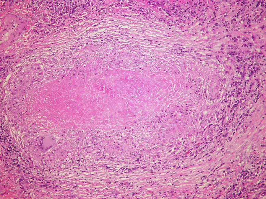
- Central caseation Structureless and granular, with no nuclei and no preserved architecture. To the naked eye it looks like soft cheese, hence the word ‘caseous’. If a stain finds bacilli, they're here.
- Epithelioid macrophages Activated macrophages with abundant cytoplasm and elongated nuclei, palisaded around the necrotic centre.
- Langhans giant cell Epithelioid macrophages fused into one large cell. Its nuclei sit in a horseshoe around the edge, leaving the centre of the cell clear.
- Lymphocyte rim Sensitised T lymphocytes at the periphery. They send the signal that activates the macrophages in the wall, so the granuloma lasts only while they're there.
- Fibroblasts and collagen Spindled cells and collagen deposited at the margin. Granulomas heal by fibrosis and calcification, and this zone later becomes the calcified nodule found incidentally at autopsy.